TREATMENT INDICATIONS
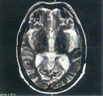
Gamma Plan ICON enables co-registration of non-stereotactic tomographic data such as MR, CT and PET to a frame-based/frame less study for the same patient. Gamma Plan ICON makes it possible to overlay the brain atlas (Schaltenbrandt & Wharen) Atlas Space on any data set. 1. Arteriovenous malformations of brain (AVMs) 2. Benign and malignant tumors, like Schwannomas or Meningiomas, Neuromas e.g. vestibular, pineal region tumors. Astrocytomas (small volume low grade) pituitary adenomas, hemangioblastomas, craniopharyngiomas, miscellaneous skull base tumors. 3. Ocular disorders- orbital tumors melanomas, eye metastasis, advanced glaucoma's, age-related macular degeneration, hemangioblastomas, angioreticulomas, pseudotumors& vegetative pain. 4. Functional disorders: Trigeminal neuralgia, Parkinson disease, other involuntary movements, intractable pain and epilepsy. 5. Adjustment therapy in the treatment of residual and recurrent lesions left unresected by conventional Neurosurgery. 6. Patients not suitable for surgery due to illness or advanced age or children. 7. In all neurosurgical cases when Gamma Knife is the only modality like thalamus and brain stem lesions.8. All hidden neck tumors are accessible for the stereotactic radiosurgery and radiotherapy. Patients return to their normal lifestyle immediately following treatment. we have with us a team of dedicated Gamma knife neuro-surgeons radio-oncologist, radio-physicist and radiologist trained in Sweden. It would be our pleasure to provide you with any information clarification that you may require.
Special Feature of Gamma Knife ICON
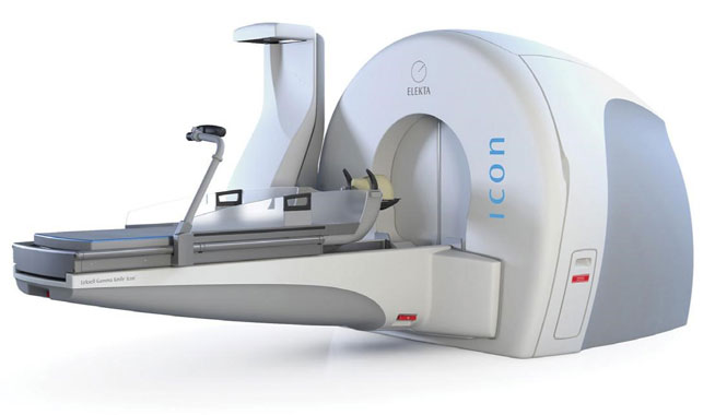
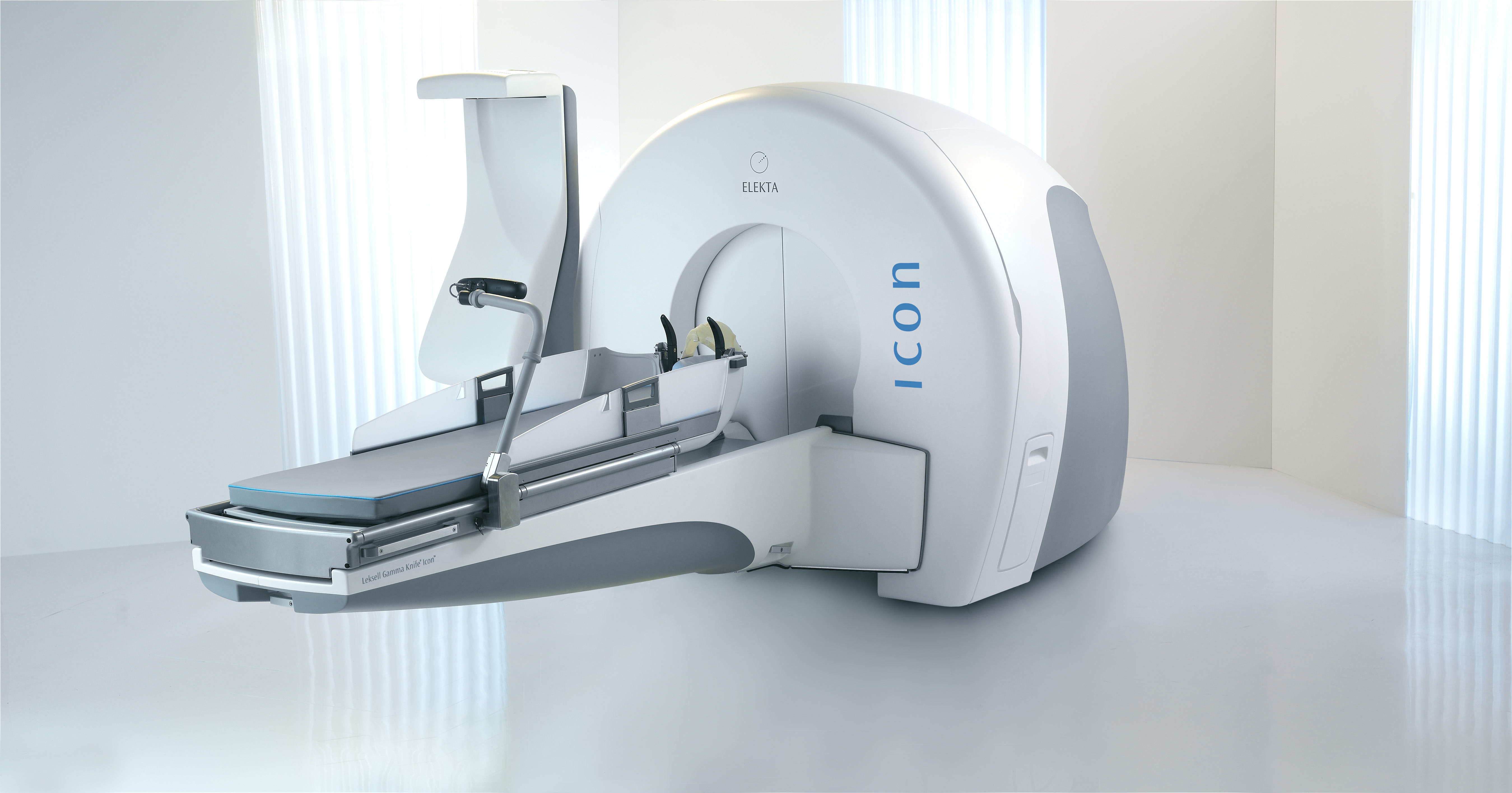
Leksell Gamma Knife® Icon™ is a dedicated radiosurgery system used in the stereotactic irradiation of intracranial structures. Surgery is achieved by delivering a prescribed dose as one or several shots of ionizing radiation to the exact site of the target. Gamma Knife radiosurgery is the most precise form of radiation therapy with an accuracy better than 0.3 mm guaranteed over life (with Platinum service contract). Icon treats a broad range of brain disorders with the lowest dose to normal tissue. Icon is equipped with integrated stereotactic Cone Beam CT (CBCT) imaging and software to continuously control the dose delivery. Icon provides several workflow options and adaptation possibilities to each individual case. The system enables multiple or single sessions, frameless and frame-based immobilization - always with the same high level of accuracy. Leksell Gamma Knife Icon introduces a number of new innovations such the following:
Adaptive DoseControl
Adaptive DoseControl™ is a comprehensive concept that confirms accuracy and ensures the precision of treatment delivery. Integrated with the control system, it gives total control through the entire process and permits real-time clinical decision-making at the time of treatment. DoseControl consists of two elements; Real-time HD Motion Management and Real Dose Delivery.
Real-time HD Motion Management
With Icon, the same precision can be achieved with frameless and frame-based immobilization. The intra-fraction motion management system monitors the patient in real time during treatment with 0.15 mm accuracy, six times better than industry standard. If the patient moves out of the pre-set threshold, the system’s gating functionality instantly blocks the radiation.
Real Rose Delivery
The unique integrated stereotactic CBCT is a new addition to Icon. Calibrated to the patient positioning system, it determines stereotactic coordinates in 3D, using bony anatomy. After co-registering the images from the CBCT and MR, the treatment plan automatically adapts to any needed correction in patient position. Thanks to the unique dose delivery attributes of Leksell Gamma Knife Icon, the system can adapt to patient rotation shot by shot, without any added mechanical uncertainty,
Online Dose Evaluation
The online Dose Evaluation enables the user to compare the dose distribution that is about to be delivered to the dose that is planned to be delivered. This is done right at the console and if needed, the plan can be adapted online quickly and easily.
Leksell GammaPlan
For Leksell Gamma Knife Icon, the seamless integration between Leksell GammaPlan®, the stereotactic CBCT, and the management system. With Leksell GammaPlan a full treatment plan can take just minutes to complete, even for complex cases. Inverse planning automatically optimizes the plan. Dose sculpting enables the precise handling of complex targets, and with Dynamic Shaping, critical structures are protected. The Convolution™ module accounts for different tissue types when the dose is being calculated.
Treatment with Leksell Gamma Knife Icon
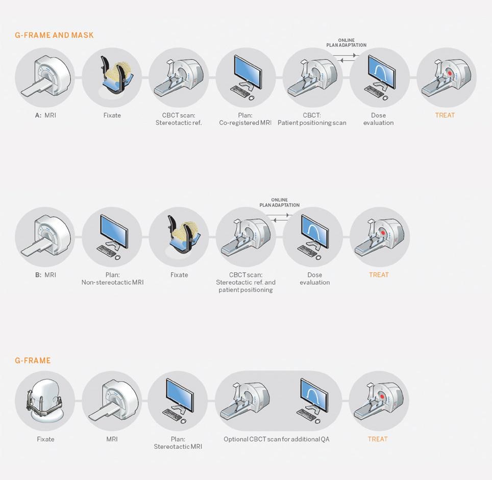
Leksell Gamma Knife® Icon™ introduces a number of workflows, either fiducial-based or reference-defined by CBCT. This brings flexibility, patient centricity, and adaptation to clinic logistics. For fiducial-based workflows, the integrated CBCT system in Icon adds an additional imaging modality, visualizing the treatment quality. The versatile workflow enables several possibilities to adapt the procedure to the clinical needs, which is based on options in patient immobilization, when to perform planning imaging and defining stereotactic reference, and on which day the tasks are performed.
Description of Leksell Gamma Knife Icon
The Leksell Gamma Knife® Icon™ system consists of several parts, physically separated into a treatment room and a control room:
• The treatment room contains the Leksell Gamma Knife® unit, which basically consists of the radiation unit, a patient positioning system, a gantry for CBCT, and a set of covers. The Real-time HD Motion Management (HDMM) system is attached to the patient positioning system. The treatment room also includes the patient surveillance system, a treatment view monitor, and a radiation warning lamp.
• The control room contains an operator console and an office cabinet. The treatment session is controlled and monitored by the operator from the control room. The control room also contains a Leksell GammaPlan® workstation. The system is electrically separated into an office side and a medical side.
• The medical side includes the Leksell Gamma Knife unit in the treatment room, and is powered and controlled by a medical cabinet, placed inside the rear cover of the radiation unit.
• The office side consists of the equipment in the control room, the patient surveillance system, the treatment view monitor, and the radiation warning lamp in the treatment room. The office side is powered and controlled by the office cabinet. The office side and the medical cabinet in the treatment room are categorized as the control systems and electronics of Leksell Gamma Knife Icon.
Radiation unit
The radiation unit is the radiation delivery system, which is made of cast iron to achieve radiation protection. It houses 192 cobalt-60 (60Co) sources and the collimator system that directs the radiation to the focus point. The sources are fixed in 8 independently movable sectors mounted on the collimator body, and contain collimators of 4, 8, and 16 mm.
Servo-controlled motors in the sector mechanism position the sources to achieve one of the conditions for Beam On (the sources are aligned with the collimators) or Beam Off (the sources are positioned and locked in the sector home position). When the treatment target is repositioned between shots, the sources are placed in the shielded sector off position between the collimators of 4 and 8 mm.
The shielding doors move horizontally to the left and right to open the radiation unit and allow the patient positioning system to move the couch to the treatment position inside the radiation cavity. A cylinder-shaped collimator cap covers the collimator holes. Glass fiber covers suspended on attachment points around the radiation unit and hanging above
Patient positioning system
The patient positioning system is the component of Icon™ that the patient reclines on for treatment. The treatment couch is automatically repositioned inside the radiation unit, moving the patient to the target coordinates as defined in the treatment plan.
The 3-axis positioning system consists of a steel framework, housing the electromechanical drive for the patient couch. An adjustable mattress is placed on the couch. The mattress has a strap that can be used to secure the patient’s arms.
The operator can maneuver the patient vertically when fitting the mask or coordinate frame (and the patient) to their respective docking device, by adjusting the position of the mattress with the manual control (one unit on each side).
Two integrated protection panels are fitted on each side of the patient couch. The panels are made of transparent plastic and can be folded up/down to facilitate easy access to the couch.
CBCT gantry
The gantry contains the parts used for CBCT.
The rotating unit, which is called the C-arm, has an X-ray tube and an image detector attached to it.
The C-arm moves the head of the X-ray tube and the image detector in a circular path around the patient.
The C-arm is attached to a tilt arm, and the other end of the tilt arm is attached to the radiation unit. The tilt arm moves the C-arm from the parked position to the scan position.
Because the gantry is mounted on the radiation unit, a fixed relation can be established between the coordinate system of the images and the radiation focus.
The complete gantry consists of the C-arm and the tilt arm fully assembled with sensors, cabling, motor, and transmission. It also includes the kV generator and the actuator for the tilt arm that are both located inside the radiation unit covers.
Real-time HD Motion Management
The HDMM system, also known as Intra-Fraction Motion Management (IFMM), is used to monitor movements of the patient during setup and treatment when immobilized by a mask fixation system (see page 20). It cannot be used when the patient is immobilized with the Leksell® Coordinate Frame.
The HDMM system is based on an infrared (IR) camera, which in combination with IR reference markers and a reflecting marker on the patient’s nose is used to detect movements. Placement of the reflecting marker on the nose is chosen because the skin on the nose is relatively thin, which gives good correlation between nose and skull movements. It is also an advantageous position for the line of sight.
A graph in the GUI shows the patient’s movements when HDMM is on. The axes of the graph shows movements in mm and duration in seconds. During treatment (gamma radiation) and when HDMM Active mode is enabled, if the patient moves out of position, the system inhibits dose delivery. If the patient moves back into position, the system resumes dose delivery after 3 seconds. In addition, if the camera cannot see the reference markers for more than 2 seconds, a treatment pause is started.
Control systems and electronics
The control systems control all the electro-mechanics of Leksell Gamma Knife Icon based on the treatment data. A comprehensive description is given in the next section.
Control Systems and Electronics
Operator console
The operator console in the control room is the interface between the operator and Leksell Gamma Knife® Icon™.
The control panel on the operator console is used to control the treatment procedure and communicate with the patient. The treatment view monitor provides a continuous display of the treatment session’s progress, including a visual display of the alarm conditions.
The mouse and the keyboard provide the user interface between the operator and the control system software that runs on the office computer, also known as the Main Computer Unit (MCU). The control system software is used to load, check, and execute the treatment as stipulated in the treatment plan exported from Leksell GammaPlan® (LGP).
The office cabinet contains the MCU, the primary LGP workstation, a network router, and an Uninterruptible Power Supply (UPS) unit for the office side of the system (Office UPS).
Patient surveillance system
A video and intercom system is used for communication with and observation of the patient.
A camera is mounted in the treatment room overlooking the patient in the treatment position. The video monitor provides continuous visual monitoring of the patient, the treatment couch, and the shielding doors of the radiation unit.
The loudspeakers and the microphone together with the TV system form the patient’s
intercom, which allows direct contact with personnel in the control room. The speakers are located in the patient couch and The different parts of the application window are the following:
The electronic control unit controls all movements of the system except for the gantry. It also delivers power to the other the microphone is close to the patient head, next to the manual control.
User interface
The control software runs on the office computer and is displayed on the treatment view monitor of the operator console.
The treatment room monitor shows a copy of the information displayed on the treatment view monitor.
There are two operating modes (versions) of the control software, a clinical mode for treatment sessions, and a service mode for service activities only. The current operating mode is clearly identified in the title bar when running the software. To preserve security, all users have to use a password at login.
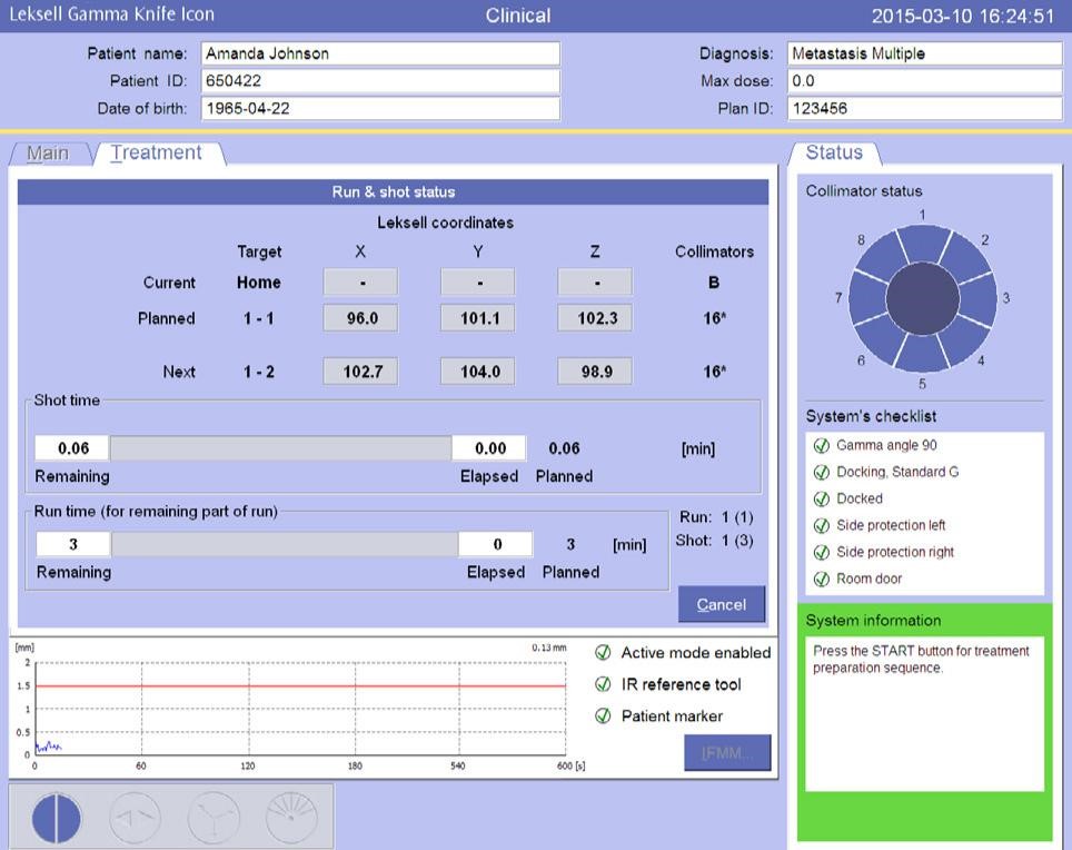
The control panel and graphical user interface together show the treatment status and the different states during the treatment flow.
On the control panel, information during the procedure is reduced to one green light and one yellow light. The green represents beam off and the yellow can alter between blinking, when there is a movement such as shielding door opening, and a steady light when beams are on.
The graphical user interface presents these states in a more detailed manner, with icons explaining the different states and movements using a yellow color in the activated icons.
• The title bar area with patient information
• The main tab area, which displays available functions
• The status tab area, which displays important system status
• The indicator area, which displays the physical status of the system
• Command buttons for the main functions.
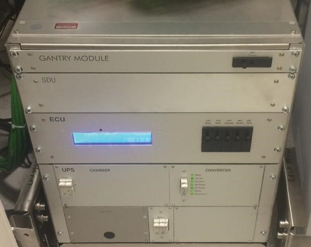
Medical cabinet
The medical cabinet is located inside the radiation cover behind the radiation unit. It contains the control electronics and the power supply system for the medical side of the system:
• The Electronic Control Unit (ECU)
• The Sector Drive Unit (SDU)
• The gantry module
• The medical UPS.
subsystems, and contains the safety system that monitors information from the sensors in the system. The sector drive unit contains one servo drive for each sector unit (8 drivers in total).
The gantry module houses the electronics that controls the movements of the gantry arms and the functions of the CBCT components.
The medical UPS, which is approved for medical use, contains the uninterruptible power supply for the system. The rechargeable batteries are maintained in a charged state by built-in chargers. In case
of failure in the mains supply, the UPS provides power for at least 20 minutes. If the duration of the power failure is longer than one minute, a treatment pause sequence is automatically initiated.
Leksell Coordinate System
The detailed graphic images of the patient’s head form the basis for treatment planning. The images can be obtained by employing one or more scanning techniques: Computerized Tomography (CT), Magnetic Resonance (MR), Positron Emission Tomography (PET), and/or angiograms (AI).
As part of the imaging process, it is essential to provide exact points of reference by means of which the shape and position of the targets can be ascertained with respect to the patient’s skull. These points of stereotactic reference provide coordinates in the Leksell® Coordinate System. MR, CT, and AI indicator boxes that are components of the Leksell Stereotactic System® can be used to impose reference fiducials during image acquisition. New to Leksell
Gamma Knife® Icon™ is the option to use the integrated CBCT instead of setting the stereotactic reference.
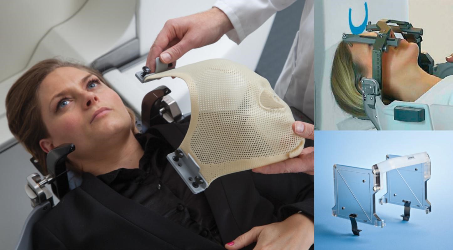
During the following radiosurgery session, the head of the patient must be entirely immobilized to maintain the accuracy of the treatment. Icon offers two methods of immobilization that provides mechanical interfaces to the docking device of the treatment equipment; frameless and frame-based fixation.
Mask fixation setup
The frameless solution includes a thermoplastic mask, a head cushion, and a mask and adapter. To give a fixation, both the mask and the head cushion are carefully shaped after the patient’s head and then hardened. For patient comfort, a knee support is used.
The patient can be undocked and re-docked using the mask immobilization. This means that the mask fixation setup can be used for fractionated treatments over the duration of a treatment period. The stereotactic reference is always set with the help of the integrated CBCT when using the mask for immobilization.
Leksell Coordinate Frame G
The frame-based solution is the Leksell® Coordinate Frame G which is the basic part of the Leksell Stereotactic System®. It is affixed to the head by means with screws and rigid corner posts. Thereby it prevents intraoperative coordinate frame displacement.
Leksell GammaPlan
Leksell GammaPlan® is sophisticated treatment planning and management software, which is fully integrated with Leksell Gamma Knife® Icon™. It provides specialized tools that make full use of the advanced technology incorporated into the system, while ensuring safe and efficient workflows.
A complete and accurate modelling of Leksell Gamma Knife Icon and patient immobilizations enable both accurate dose calculations and simulation of all geometries to ensure safety in treatment delivery. A range of highly responsive forward and inverse planning tools, tailored for the systems unique dose characteristics, allows for interactive dose sculpting, and makes it possible to complete plans in minutes, even for complex and multiple targets. Convolution™ provides optional heterogeneity correction for targets near bone and air cavities. Both manual and automatically calculated dose statistics are available for efficient plan review.
Leksell GammaPlan supports several workflows for treatment planning and delivery with Icon. For frameless treatments, a fully integrated and guided workflow is provided for automatic correction and online evaluation of delivery dose based on the current patient position from CBCT images. For frame-based treatments, CBCT may be used as an optional quality assurance for patient positioning. Re-planning tools with optional dose summation are provided to facilitate plan adaptation for both single-session and fractionated treatments. Re-treatment functionality provides tools for the efficient management of reoccurring disease, in particular metastases. Pre-planning makes it possible to plan on non-stereotactic images.
Leksell GammaPlan supports DICOM image studies obtained from MR, CT, CBCT, and PET scans as well as projective X-ray angiograms. Stereotactic Leksell® coordinates are obtained by localizing image studies acquired with Leksell Coordinate Frame G, or stereotactic CBCT on Leksell Gamma Knife Icon. Non-stereotactic image studies, such as frameless MR, PET, and MEG data images can be co-registered and fused to other image studies for the same patient based on Mutual Information in the images. The patient skull shape can be automatically segmented from MR or CT images, and regions of interests can be delineated on both tomographic images and projective X-ray angiograms. The fully configurable image workspace allows for simultaneous display and exploration of multiple image studies while planning.
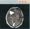
Functional planning is supported with the optional AtlasSpace™ (Schaltenbrand & Wahren) brain atlas, and functional targeting can be done based on the Relative Intercommisural line.
Thanks to a client-server architecture, multiple LGP workstations have instant access to all patient data recorded in the online database, shared with Leksell Gamma Knife Icon. Connectivity with external systems is provided via standard DICOM interfaces. DICOM images and DICOM RT Structure Sets can be both imported from, and exported to external systems over the network. It is also possible to export DICOMRT Dose.
Leksell GammaPlan add-ons
• Inverse Planning – Highly responsive conformity-based optimization
• Convolution – Increased dose accuracy near heterogeneities
• DICOM RT – Support for increased connectivity
• MOSAIQ® Connectivity (optional) – See below.
surgical draft planning and post-operative follow-up by co-registering image studies from DICOM, CT, MR, PET, and MEG
• WarpSpeed™ – Real-time update of dose distribution
• ImageMerge™ – Use frameless images. Any non-stereotactic image study can be matched with a Leksell® Coordinate Frame scanned image.
• ColorPET™ – Support for color PET images
• Functional planning – Relative Intercommisural line and functional targets
• AtlasSpace (optional) – Schaltenbrand & Wahren brain atlas.
MOSAIQ Connectivity
The optional MOSAIQ Connectivity provides seamless integration with MOSAIQ Oncology Information Management System for complete patient charting and a streamlined clinical workflow. The complete picture of any Icon patient case is accessible from any MOSAIQ workstation in the hospital. In addition to importing into MOSAIQ treatment plans and treatment records generated by Icon, clinical users can automate the billing and scheduling processes, and save valuable time and resources.
Radiation unit
The following section describes selected components within the radiation unit.
All components are chosen from well-recognized suppliers to secure reliability and to optimize the performance of Leksell Gamma Knife® Icon™.
Patient positioning system
The following section describes selected components within the patient positioning system.
All components are chosen from well-recognized suppliers to secure reliability and to optimize the performance of the system.
CBCT gantry
The following section describes selected components within the gantry.
All components are chosen from well-recognized suppliers to secure reliability and to optimize the performance of the system.
Real-Time HD Motion Management
Optical tracking system with frame-based reference markers and a tracking marker on the patient’s nose. Foldable arm that allows the tracking camera to be moved to a parked position when not used.
Control systems and electronics
Office cabinet
The office cabinet is a standard 19” cabinet that holds the MCU, the office UPS, a network router, and the primary LGP workstation.
Main Control Unit
The Main Control Unit (MCU), or office computer, is an industrial grade rack PC. The patient’s treatment plan is sent from the LGP database to the MCU via the network router. The MCU then communicates to the ECU the part of the treatment that is to be performed, and updates the treatment status after each performed part.
In addition, the MCU monitors the status of the Icon system and the office UPS.
Office Uninterruptible Power Supply
The office UPS is a standard rack battery backup powered by the mains inlet. It produces the same alternating voltage on its outputs as on the input (and the same frequency).
The office UPS comes in different configurations to meet country-specific requirements.
Network router
The Ethernet gateway provides secure and reliable access to the database. It also delivers secure and reliable high-speed interconnection between the different subsystems of Leksell Gamma Knife Icon.
LGP workstation
The primary LGP workstation is a HP Z620 or better, with at least 32GB memory.
Note: Although the primary Leksell GammaPlan® workstation is shipped with Leksell Gamma Knife Icon, the LGP workstation (as well as optional external units) has its own registration.
Operator console
Connection and Isolation Unit
The Connection and Isolation Unit (CIU) ensures that all interconnections between the office and the medical sides (safety signals and the CAN bus to the ECU) respect the required isolation.
The CIU also handles the mains power for the office side and distributes the interruptible power (via Power On control) to the devices that can be shut down (mainly the monitors).
It finally takes care of the indicators and the buttons on the operator panel.
The following signals are accessible via relay contacts from the CIU on the back panel of the console:
• Emergency alarm
• Radiation On (not Beam Off)
• In treatment (Beam On).
Patient Surveillance System
The Patient Surveillance System (PSS) is included in the operator console as a separate unit and handles the video/audio signals of the Icon system.
It is possible to connect a video recorder on the ‘auxiliary’ outputs. External audio systems can be connected and played over the sound system integrated in the patient positioning system.
Medical cabinet
The medical cabinet contains the SDU, ECU, Medical UPS, and gantry module. It is an Elekta-made cabinet to fit the shape of the back of the radiation unit.
Sector Drive Unit
The Sector Drive Unit (SDU) contains 8 servo drivers (one for each sector). When the servo drivers are disabled (either by the ECU or in case of Emergency Stop or Emergency Exit), the sector motors are completely disconnected from the drivers.
In case of Emergency (Stop or Exit), a fixed voltage is applied directly to the sector motors pulling them back to a locked shielded position.
Electronic Control Unit
The Electronic Control Unit (ECU) consists of two complete Power PCs (PPC) with peripherals (RAM, ROM, inputs, outputs, and CAN interfaces).
PPC1 controls all movements of the Icon system via CAN messages to the other subsystems. PPC2 monitors all CAN traffic and the PPC1 commands. If any failure or error is detected, the PPC2 activates the safety system.
In order to always display the planned and elapsed treatment times, the ECU cannot be shut down by the MCU. Nevertheless, in power off mode the microprocessors are reset and all outputs are inactivated.
The FPGA (field programmable gate array) handles the reading of the redundant scales of all moving axis and makes this information available to both PPC1 and PPC2. In the FPGA, an independent timer measures the Beam On time.
The treatment time is read by the PPCs. PPC1 decides when to stop the treatment and if it fails, PPC2 will terminate the treatment. Information from both PPC1 and PPC2 are shown on the ECU display. Typical information is treatment time and run number.
The following signals are accessible from the back panel of the ECU:
Declaration of conformity
Leksell Gamma Knife Icon complies with the requirements of the Medical Device Directive 93/42/EEC. The product is CE marked.
Standards approval
EN/IEC 60601-1
Medical electrical equipment, Part 1: General requirements for safety.
EN/IEC 60601-1-2
Collateral standard: Electromagnetic compatibility.
IEC 60601-2-11
Medical electrical equipment, Part 2: Particular requirements for the safety of gamma beam therapy equipment.
AAMI ES60601-1
Medical electrical equipment: General requirements for safety.
C22.2 No 601.1:08
Medical Electrical Equipment, Part 1: General Requirements for safety.
IEC 60601-1-3
Radiation protection in diagnostic X-ray equipment.
IEC 60601-2-28
Particular requirements for the basic safety and essential performance of X-ray tube assemblies for medical diagnosis.
IEC 60601-2-68
Particular requirements for basic safety and essential performance of X-ray based Image Guided Radiotherapy Equipment for use with electron accelerators, light ion beam therapy systems and radionuclide beam therapy systems.
• External Pause
• External Emergency Stop
• Treatment room door sensor.
Medical Uninterruptible Power Supply
The medical Uninterruptible Power Supply (UPS) delivers the 24 and 48 V DC needed for the Icon system. It is approved for medical use.
Alarms are generated if the converter modules fail, if the battery fails or if the internal temperature rises above a certain limit.
The UPS allows a 20-minute stand-by at mains failure.
Gantry module
IEC classification
Leksell Gamma Knife Icon is classified as follows:
IEC 60601-1 Type of protection against electric shock IEC 60601-1 Degree of protection against electric shock
IEC 60601-1 Methods of disinfection recommended by the manufacturer
IEC 60601-1 Degree of application safety in the presence of flammable anesthetic mixture with air or with oxygen, or with nitrous oxide
Class I equipment
Type B applied parts
Disinfectable equipment (or elements).
Equipment NOT suitable for use in the presence of a flammable anesthetic mixture with air or with oxygen, or nitrous oxide.
The gantry module acts as a bridge for the ECU to control the movements and check the sensors of the CBCT. It also allows communication between the MCU and the kV generator and handles the signal for exposures with respect for safety. During CBCT, the gantry module saves the exposure angles in real time and checks that each picture has been exposed. This information is then transmitted to the MCU for reconstruction.
Modifications
Elekta reserves the right to make changes to the product specifications and equipment covered thereby, especially where such changes are anticipated to improve performance of the product or parts, or are required to satisfy the product specifications, or are expected to improve reliability, quality or the value of the product.
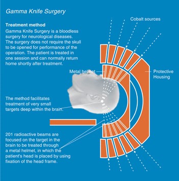
Gamma Knife® surgery is a well-established treatment method used to treat selected targets in the brain. Leksell Gamma Knife® is not a knife in normal sense of the word. The doctor makes no incisions in your head. Instead, very precisely focus beams of radiation directed to the treatment area in the brain. Gamma Knife® surgery offers a safe and effective treatment for more than 50,000 patients every year. The treatment procedure is simple, painless and straightforward. The treatment consists of four steps:
Gamma Knife® surgery is a unique method that delivers extremely focused radiation beams to targets in the brain. 201 individual beams converge to one focal point. The radiation source used is called cobalt. These sources are positioned in a hemisphere so that all the beams can converge on a single point. The shape and dose of the radiation is optimized to hit only the target, without damaging surrounding healthy tissue.
- Painless
- Bloodless
- Precise
- Low-cost
- Experiencedteam
- Dedicatedhospital
- Day care treatment
Before the Treatment
Before treatment your doctor will inform you about the entire procedure. Gamma Knife® surgery does not require cutting or shaving of your hair. The next step is the application of the head frame.
The Head Frame
A key component in Gamma Knife® surgery is the stereotactic head frame. The frame allows the doctor to accurately pinpoint the target to be treated in your brain. This lightweight frame, which is attached to your head with four screws, ensures that the radiation beams can be directed with precision to the target. The frame also prevents your head from moving during imaging and treatment procedures. Local anesthetic agent is applied where the screws are to be fixed.
Imaging
After the head frame is in place, it is time for imaging to be done; such as magnetic resonance imaging (MRI), computed tomography (CT) or angiography. Imaging is required to determine the exact size, shape and position of the target in the brain. During imaging, a coordinate box is placed on the head frame to provide reference points on the images for the treatment plan. After imaging, the coordinate box is removed.
Treatment Planning
Once your images have been taken, you can rest while your physician develops a very precise and accurate treatment plan. No two treatment plans are alike; every patient's plan is individually designed to address the specific medical condition. The doctor, very often together with another specialist in the team, makes the plan in a specially designed computer and calculates how the treatment should be performed. This usually takes a couple of hours.
The Execution of Treatment
Once your treatment plan is completed, the actual treatment can start. You will lay down on the treatment couch and the head frame will be attached to the helmet. You are awake during the procedure and will be able to communicate with your doctor or nurse through an audio and video connection. When the treatment begins, the couch will move into the dome section of the unit. The treatment is silent and totally painless. Often you will be able to listen to music or recitation of holy Quran during the treatment. The team will be monitoring the procedure at all times. The treatment will last a few minutes to half an hour, depending on the size and shape of the target.
After the Treatment
When your treatment is complete, the head frame will be removed. If you had an angiogram, you might have to lay quietly for several more hours. Some patients experience a mild headache or minor swelling where the head frame was attached, but most report no problems. Your doctor will tell you whether or not he wants you to stay overnight for observation or if you can go horse immediately. Either way, you should be able to return to your normal routines in another day or so.
Follow-up
The effects of your treatment will occur over time. Radiation treatments are designed to stop the growth of tumors or lesions, which means that the effect will be seen over a period of weeks or months. Your doctor will stay in contact with you to assess your progress, which may include follow-up MRI, CT or angiography images at interval of 3, 6, 12, 18 or 24 months.